HIGHLY MULTIPLEXED SPATIAL PROTEOMICS FOR TRANSLATIONAL RESEARCH
High-Resolution Spatial Proteomics Accelerates Personalized Insights Into Cancer
Leverage our expertise and the power of MIBI to gain deeper understanding of the tumor microenvironment


Find out how MIBI-enabled spatial proteomics can provide more detailed insights into the tumor microenvironment
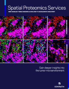
Through its Spatial Proteomics Services, Ionpath’s expert team can help you obtain actionable insights by using MIBI high-resolution spatial proteomics to image and analyze your precious tissue samples.
MIBI-based analysis provides information—not available with IHC—that many researchers have utilized to unravel novel understanding about the tumor microenvironment in several cancers and disease systems (see MIBI publications).
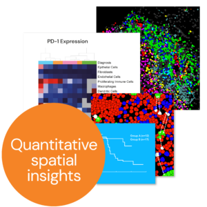
- Map and quantify cell populutations within the tumor microenvironment
- Quantify expression of immune checkpoints and other protein biomarkers with spatial context
- Profile, visualize and map the immune infiltrate and tumor-immune boundaries

MIBI reveals actionable insights
MIBI analysis of breast cancer tissue. A: MIBI image of a DCIS tumor and the resulting spatial phenotype map. B: Comparison of the primary DCIS diagnosis (left) and invasive recurrence (right) using MIBI imaging revealed structural and funcutional coordination in the tumor stroma correlated with disease progression. (Risom et al. Cell 2022)
See how we can help accelerate your research.
Leverage our expertise.
Want to talk with an expert now?
Contact us at mibi@ionpath.com