MIBIplus
Unlocking Spatial Proteomics with Precision

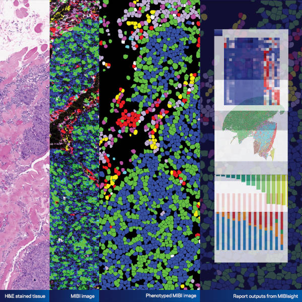
With MIBIplus, Ionpath offers an all-in-one, single-cell spatial proteomics solution that lets you detect over 50 cell phenotypes in a single scan. Our end-to-end service supports projects of all sizes, at any frequency, and at every stage of your development program so you can confidently advance your research.
Ionpath’s groundbreaking MIBIscope™ 3.0 is at the core of MIBIplus, designed to maximize the data density of your precious tissue samples. Engineered to capture spatial data with unparalleled precision, it detects high, medium, and low protein expressions simultaneously, all while preserving the tissue’s spatial architecture.
Our expert technical team analyzes your samples and delivers comprehensive, actionable insights through detailed reports. These reports are customizable, allowing you to tailor results to your specific research needs. With our interactive analysis tools, you can revisit, reanalyze, and regenerate reports with just a few clicks, enabling continuous discovery.
MIBIplus Panels: Comprehensive and Customizable
MIBIplus features an expanded suite of both ready-to-use and customizable panels, purpose-built to answer your key research questions:
Application Areas
- Immunology: Leverage the power of spatial proteomics to advance immunology research. This panel includes 27 key markers, such as Granzyme B, Ki-67, ICOS, IDO-1, and B7-H3, alongside 3 advanced checkpoint markers, enabling a detailed assessment of immune cell infiltration, activation states, and checkpoint expression. Ideal for studying immune responses, characterizing immune cell interactions, and exploring mechanisms of immune regulation.
- Tumor Immune Landscape: Characterize immune cell phenotypes and functional states within the tumor microenvironment, assess immune activity, and compare pre- and post-treatment samples. Key markers include CD3, CD8, PD-L1, and FOXP3, enabling a comprehensive understanding of immune dynamics.
- Hot vs. Cold Tumor: Distinguish immune-responsive (hot) tumors from immune-deserted (cold) tumors and evaluate the immune landscape driving tumor progression or regression. This panel includes checkpoints like PD-1 and CTLA-4, along with markers for T-cell activation and suppression.
- Inflammation Panel: Uncover the cellular and molecular drivers of inflammation in both healthy and diseased tissues. Target key immune cells like macrophages, neutrophils, and T cells, along with cytokines such as IL-1β, IL-6, TNF-α, and IL-10, to assess inflammatory states and their resolution. This panel supports research in autoimmune diseases, chronic inflammation, fibrosis, and the tumor microenvironment.
MIBIplus Panel Suite
MIBIplus panel are a versatile collection of ready-to-use and customizable panels designed to deliver precise insights and empower your spatial biology research.
General/Structural Panel: This foundational assay is designed to provide deep insights into tissue architecture and cellular functions. It includes key markers such as:
- CD31: For vascular and endothelial cell analysis.
- Podoplanin: A marker for lymphatic cells.
- Ki-67: To assess cell proliferation and activity.
- α-SMA and Vimentin: For fibroblast and smooth muscle phenotyping.
- Histone H3 + dsDNA: To examine nuclear structure and DNA content.
This panel serves as a robust starting point for spatial analysis, enabling researchers to customize their studies further by adding specific antibodies or integrating additional subpanels focused on areas like immune profiling, cellular metabolism, or tumor microenvironment characterization.
New Panels Coming Soon!
Exciting advancements are on the horizon for the MIBIplus panel suite. Launching in Q1 2025, these new panels will expand your research capabilities with cutting-edge tools designed to profile diverse cellular phenotypes and markers, such as:
- Fibroblasts, myofibroblasts, and smooth muscle
- Immune, lymphatic, and endothelial cells
- Key markers like CD45, Keratin, Podoplanin, and HL1 ABC + Na/K ATPase

Flexibility to Expand Your Insights
MIBIplus doesn’t just stop at delivering actionable results. If additional markers are required after receiving your report, our technology enables you to revisit and rescan the same tissue sample, adding new markers without compromising the integrity of the original data. This flexibility ensures that your research keeps pace with evolving questions and discoveries.
Need something specific? Our customizable panel options allow you to add markers tailored to your unique research requirements.
MIBIplus: Transforming Tissue Analysis
MIBIplus enables unparalleled insights into tissue biology, empowering researchers to:
- Identify distinct immune cell types and their spatial distribution.
- Analyze immune activity and functionality within tumor samples.
- Distinguish responder versus non-responder phenotypes.
- Define unique spatial signatures across treatment groups.
- Reveal mechanisms of action and unlock novel insights into cancer immunobiology.


Project Features
- Simultaneous measurement of 50+ cell phenotypes and tissue morphology from a single tissue slide.
- Comprehensive spatial analysis reports with customizable features.
- Access to MIBIplus Manager for interactive data visualization, reanalysis, and report regeneration.
- Ready-to-use panels, including:
- Human Immuno-Oncology Panel (27 markers + 3 advanced checkpoint
markers). - Tumor Immune Landscape Panel.
- Cellular Metabolism Panel.
- Hot vs. Cold Tumor Panel.
- Inflammation Panel.
- Human Immuno-Oncology Panel (27 markers + 3 advanced checkpoint
- Support for FFPE tissues (excluding de-calcified tissue).
- Coming Soon: Mouse Immuno-Oncology Panel.
Deliverables
- Single-cell segmented and phenotypic dataset in CSV format.
- Study-level summaries with key quantified insights.
- Automated standard reports with visual and statistical outputs.
- Full access to MIBIplus Manager for dynamic data visualization and iterative discovery.
- Flexibility to rescan tissue samples, enabling additional markers to be added as your research questions evolve.
Discover more with MIBIplus — your key to unlocking deeper spatial proteomics insights
and accelerating your research breakthroughs.
