MIBI Technology
MIBI™ technology (Multiplexed Ion Beam Imaging) uses Secondary-Ion Mass Spectrometry (SIMS), a type of mass spectrometry traditionally used in the semiconductor industry, to image antibodies tagged with monoisotopic metal reporters.
This unique technology enables:
- Visualization of 40+ markers simultaneously
- Imaging at the sub-cellular resolution
- Detection of low abundance proteins
- Rescanning of slides at multiple resolutions
Workflow
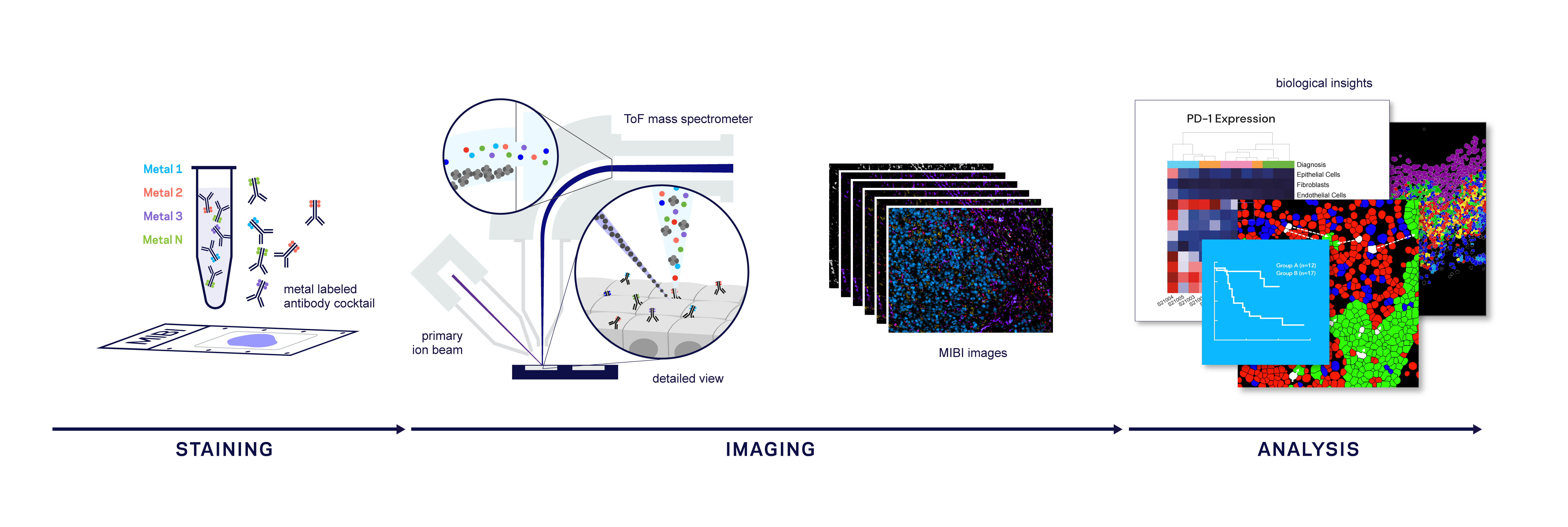
The system uses a simple, streamlined workflow that adapts easily for both pathology and clinical imaging labs. Samples are prepared using a traditional IHC protocol and data are collected in less than 1 hour using the MIBIscope™.
- STAINING FFPE or frozen tissue is stained with antibodies, each labeled with a unique metal isotope. Each antibody binds to a single protein target.
- IMAGING An ion beam is scanned across the tissue releasing secondary metal isotope ions which are detected and quantified by a ToF mass spectrometer.
- ANALYSIS Bioinformatic analysis and access to cloud-based MIBItracker Software for easy data visualization.
Highly Multiplexed Tissue Imaging
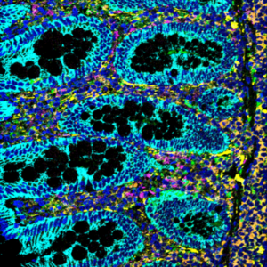
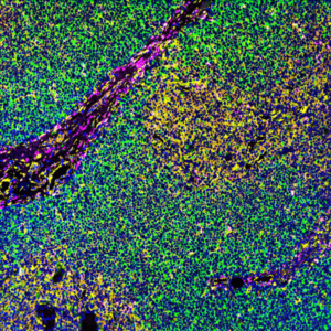
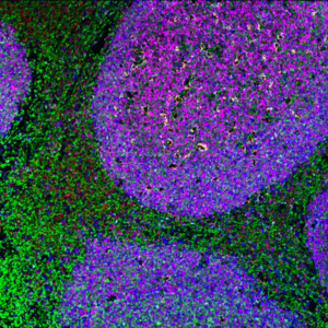
In a single scan, the MIBIscope can measure expression of 40+ targets including both high and low abundance proteins at light microscopy resolution.High Sensitivity
Low abundance proteins such as PD-1 and FOXP3 that can be missed using other techniques are easily detected with the MIBIscope. With no probe background or autofluorescence to interfere with weak signals, the high sensitivity that results ensures all high, medium and low-abundance targets are detected in the same scan.True Sub-Cellular Resolution
With resolution comparable to light microscopy systems, MIBIscope system lets you analyze and interpret cellular interactions. Cellular structures as small as 250 nm can be imaged, enabling downstream cell segmentation data analysis. Because the MIBIscope doesn’t fully ablate the sample, a survey scan can be done to select specific regions of interest followed by high-resolution imaging.Rescan Slides at Multiple Resolutions
The system’s imaging workflow enables scientists and pathologists to scan a whole tissue section faster at low resolution, then rescan regions of interest at high resolution. Precious tissue is conserved and may be scanned repetitively.

