Immuno-oncology
One Image. All the Answers
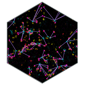
With MIBIscope™, scientists can analyze multiple markers simultaneously using a single slide. Expression can be measured at the single-cell level with subcellular resolution while preserving spatial information.
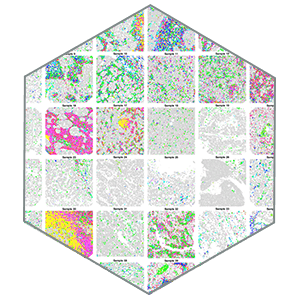
In a recent study published in Cell, A structured tumor-immune microenvironment in triple negative breast cancer revealed by multiplexed ion beam imaging, Keren et al. used MIBI technology to comprehensively profile the tumor microenvironment of 41 triple-negative breast cancer patients. The authors simultaneously imaged 36 proteins at a subcellular resolution in diagnostic surgical pathology samples.
Using a multi-step analysis, including cell segmentation, single-cell analysis and spatial analysis, the authors revealed how tumor expression and immune composition are interrelated within tissue context, and how this correlates with overall survival in TNBC. The authors showed how the MIBI™ multiplexed imaging approach can be used to stratify patient populations and identify a unique patient population that will respond to therapy.
Dr. Margaret Shipp
Professor of Medicine and Director Lymphoma Program
Dana-Farber Cancer Institute
Learn more about the publication


A Structured Tumor-Immune Microenvironment in Triple Negative Breast Cancer Revealed by Multiplexed Ion Beam Imaging
Keren et al.
Cell. 2018 Sep 6;174(6):1373-1387
Webinar presenting the publication data: Comprehensive Enumeration of Immune Cells in Solid Tumors
Michael Angelo, PM, PhD
Assistant Professor of Pathology. Stanford Medicine, Dept. of Pathology
Visualize the 41 samples in the Cell publication with MIBItracker software. Then export the data for analysis using our analysis workflow.