CD3 Antibody [MRQ-39] – 159Tb


![CD3-MRQ39-IHC-staining-FFPE-human-thymus CD3-[MRQ39]-IHC-staining-FFPE-human-thymus](https://www.ionpath.com/wp-content/uploads/2020/11/CD3-MRQ39-IHC-staining-FFPE-human-thymus.png)

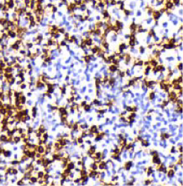
Background: CD3 is a pan-T-cell marker that reacts with an antigen present at the surface and cytoplasm of both immature and mature T lymphocytes. CD3 is also expressed in almost all T-cell lymphomas and leukaemias, and can therefore be used to distinguish them from B-cell and myeloid neoplasms.
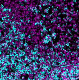
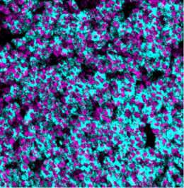
Validation: Each lot of conjugated antibody is quality control tested by staining tissue following the MIBI Staining Protocol optimized for the applicable tissue format with subsequent MIBIscope analysis using the appropriate positive and negative tissue field of views. These results are pathologist verified.
Recommended Usage: Human FFPE: 1:100 dilution.
For optimal results, the antibody should be titrated for each desired application.
References
Leong AS, Cooper K, Leong FJ (2003). Manual of Diagnostic Cytology (2nd ed.). Greenwich Medical Media, Ltd. pp. 63–64. ISBN 1-84110-100-1.
* Conjugate tested on human tissue.