CD3e Antibody – 159Tb

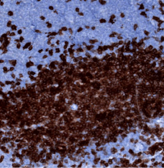
IHC: CD3e staining of FFPE mouse spleen

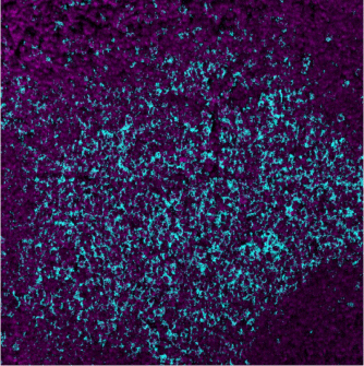
MIBI: CD3e staining (cyan) of FFPE mouse spleen, costained with dsDNA (magenta)
Background: CD3-epsilon (CD3e), together with other CD3 subunits, forms the CD3-TCR complex. It is used as a pan-T-cell marker that reacts with an antigen present at the surface and cytoplasm of both immature and mature T lymphocytes, including NK/T cells. CD3e is also expressed in almost all T-cell lymphomas and leukaemias, and can therefore be used to distinguish them from B-cell and myeloid neoplasms.
Validation: Each lot of conjugated antibody is quality control tested by staining tissue following the MIBI Staining Protocol optimized for the applicable tissue format with subsequent MIBIscope analysis using the appropriate positive and negative tissue field of views.
Recommended Usage: Mouse FFPE: 3.5 ug/mL dilution.
For optimal results, the antibody should be titrated for each desired application.
MIBI technology: Learn more about MIBI™ Technology, a multiplex IHC technology with unmatched sensitivity and true subcellular resolution.
References
Leong AS, Cooper K, Leong FJ (2003). Manual of Diagnostic Cytology (2nd ed.). Greenwich Medical Media, Ltd. pp. 63–64. ISBN 1-84110-100-1.
* Conjugate tested on mouse FFPE tissue.