MIBI Case Study | Breast Cancer
Breast Cancer: MIBI Spatial Proteomic Signatures of Progression
Ductal carcinoma in situ (DCIS) is a pre-invasive lesion that is thought to be a precursor to invasive breast cancer (IBC). DCIS comprises approximately 20% of newly diagnosed breast cancer cases, and unlike IBC, is not life-threatening. However, if left untreated, up to half of DCIS patients will develop IBC within 10 years, so clinical management has trended towards presuming all patients are progressors and treating them with surgery, radiation therapy, and pharmacological interventions. Thus, understanding the central biological features in DCIS that drive the transition to IBC is a critical unmet need for guiding appropriate patient care.
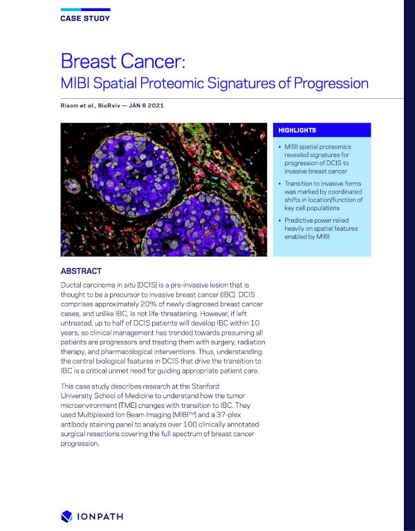
This case study describes research from Stanford University School of Medicine, Washington University School of Medicine, Duke University and Arizona State University, to understand how the tumor microenvironment (TME) changes with transition to IBC. They used Multiplexed Ion Beam Imaging (MIBI™) and a 37-plex antibody staining panel to analyze over 79 clinically annotated surgical resections covering the full spectrum of breast cancer progression.

Content
- MIBI spatial proteomics revealed signatures for progression of DCIS to invasive breast cancer
- Transition to invasive forms was marked by coordinated shifts in location/function of key cell populations
- Predictive power relied heavily on spatial features enabled by MIBI