Case Studies
MIBI Case Studies
Discover a variety of applications for comprehensive single-cell analysis, with spatial context—all in one image with multiplexed ion beam imaging (MIBI) technology.

MIBI Case Study | Diffuse Large B-Cell Lymphoma
Uncovering Insights in Diffuse Large B-Cell Lymphoma with MIBIscope™
Using the MIBI technology, the team at BMS systematically characterized cellular neighborhood clusters (CNCs) and their spatial patterns, linking these findings to clinical outcomes and enhancing the understanding of DLBCL at an unprecedented level. It allowed the researchers to visualize and quantify the spatial relationships between tumor and infiltrating non-tumor cells providing a richer context than bulk transcriptomic data alone.

MIBI Case Study | Cancer Treatment
Cancer Treatment: MIBI Spatial Proteomic Signatures of RBN-2397’s Efficacy
This study highlights RBN-2397 as a promising candidate for cancer immunotherapy. Leveraging Ionpath’s MIBI™ technology, researchers gained valuable insights into the drug’s ability to enhance immune responses within the TME, paving the way for improved cancer treatment strategies and better patient outcomes. MIBI™ technology was crucial in enabling simultaneous detection of multiple immune cell types, quantifying immune cell changes between baseline and treatment, and providing high-resolution images to visualize immune cell positions relative to tumor cells.

MIBI Case Study | Breast Cancer
Breast Cancer: MIBI Spatial Proteomic Signatures of Progression
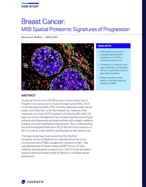
This case study describes research from Stanford University School of Medicine, Washington University School of Medicine, Duke University and Arizona State University, to understand how the tumor microenvironment (TME) changes with transition to IBC. They used Multiplexed Ion Beam Imaging (MIBI™) and a 37-plex antibody staining panel to analyze over 79 clinically annotated surgical resections covering the full spectrum of breast cancer progression.