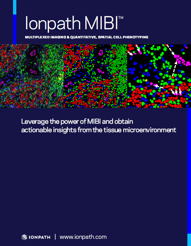
HIGHLY MULTIPLEXED SPATIAL PROTEOMICS FOR TRANSLATIONAL RESEARCH
Novel Discoveries in Spatial Biology with MIBI
Join the researchers already leveraging the high image quality and high-plex tissue imaging of MIBI

Learn how Ionpath MIBI imaging has been enabling advances in translational research and drug development
- MIBI delivers superior image quality to generate quantitative spatial phenotype mapping of the tissue microenvironment with very high resolution
- MIBI delivers rich tissue information in detail: identification and enumeration of cell populations; quantification of protein expression (e.g., checkpoint proteins); and spatial interaction insights (e.g., immune infiltration and tumor-immune boundary mapping)
- MIBI provides high multiplexing with a streamlined, efficient workflow—just a single staining step and a single imaging step
- MIBI in action. MIBI has already been used in drug development studies and for novel discovery in immuno-oncology, neuroscience, and infectious disease research (see MIBI Publications)

MIBI reveals actionable insights
IHC and MIBI image and data series. MIBI data from a head and neck cancer study that revealed infiltration by activated cytotoxic T cells in a subset of patients treated with a neoadjuvant PD-1 inhibitor therapy.
IHC and MIBI image and data series. MIBI data from a head and neck cancer study that revealed infiltration by activated cytotoxic T cells in a subset of patients treated with a neoadjuvant PD-1 inhibitor therapy.
Want to talk with an expert now?
Contact us at mibi@ionpath.com